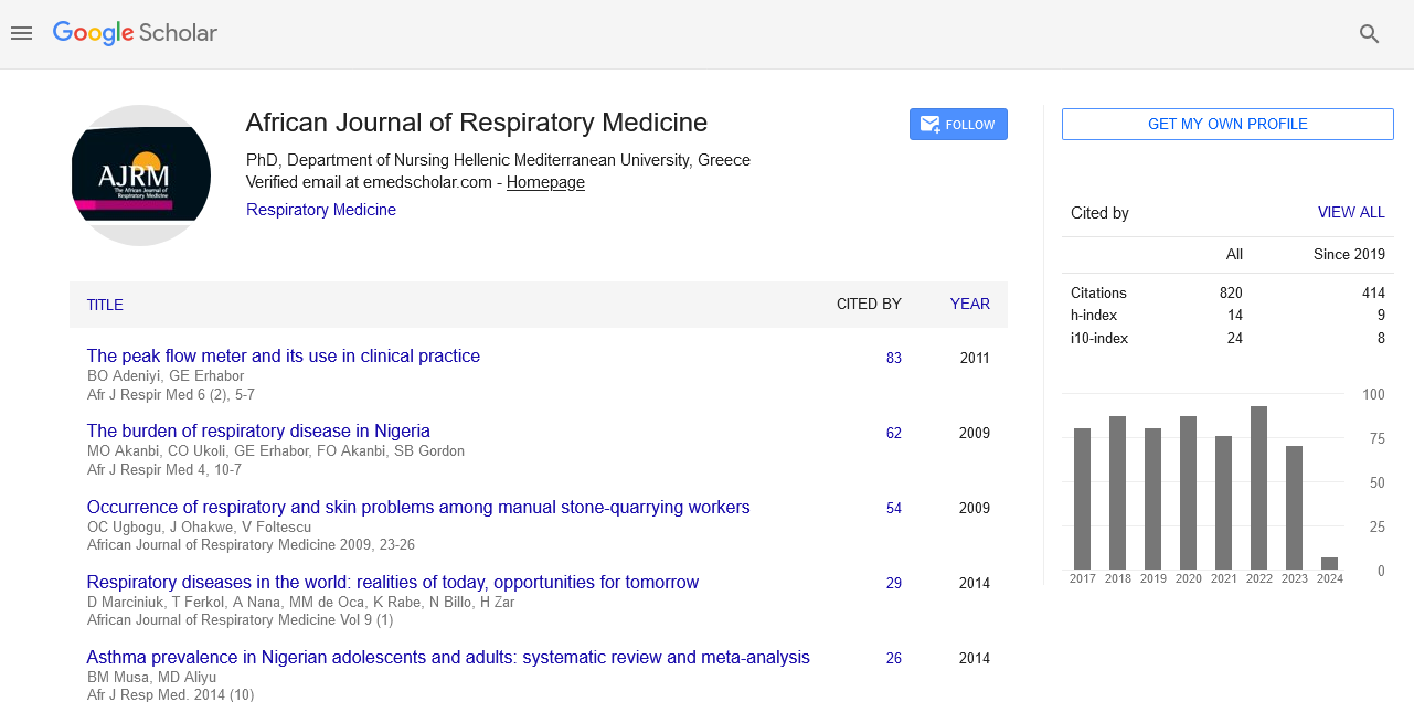
African Journal of Respiratory Medicine received 855 citations as per google scholar report
Research - (2021) Volume 0, Issue 0
Published: 28-Sep-2021
The co-infection of severe acute respiratory syndrome coronavirus 2 (SARS-CoV-2) infections with the bacterial disease has been reported from the other regions of Iran, and Abadan is no exception. Atypical pathogens infections can cause a major burden on health and treatment services. Also, they may share similar radiographic findings and clinical features with SARS-CoV-2 that making a thorough differential diagnosis essential. In this study, we have found ten cases of a patient diagnosed with Mycoplasma pneumoniae (M. pneumoniae) and Legionella pneumophila (L. pneumophila), and concomitant SARS-CoV-2 pneumonia, highlighting the need for an etiological investigation of this pandemic.
SARS-CoV-2; COVID-19; M. pneumoniae; L. pneumophila; C. pneumonia
The pandemic coronavirus disease 2019 (COVID-19), caused by Severe Acute Respiratory Syndrome Coronavirus 2 (SARS-CoV-2), was originated from Wuhan, Hubei Province, China in December 2019.1,2 It rapidly spread worldwide and became a global threat.3 The major route of transmission for SARS-CoV-2 infection is the delivery of respiratory droplets from symptomatic and asymptomatic individuals to healthy people.4 Previous studies on respiratory infection ensure that co-infections with a diversity of co-pathogens can occur.5 Furthermore, co‐infections and superinfections are common complications in respiratory viral infections. Some viral infection‐related bacterial species include Staphylococcus, Streptococcus pneumoniae, Enterococcus, Klebsiella pneumoniae, Enterobacter, Escherichia coli, Legionella pneumophila, Acinetobacter, Mycoplasma pneumoniae, Chlamydia pneumonia, and Pseudomonas.6-8We aimed to assess the rate of bacterial and SARS-CoV-2 co-infections and their outcomes among COVID-19 patients are living in Abadan, Iran.
Sample collection
We have collected respiratory samples from forty RT-PCRconfirmed COVID-19 cases with upper respiratory system symptoms, which live in the city of ABADAN, after patients’ approval for participating in the study. The inclusion criteria are mild, moderate, and severe symptoms and patients with ARDS. All of clinical and laboratory manifestations have been collected carefully.
Detection of Community-Acquired Pneumonia (CAP) in COVID-19 patients
We used polymerase chain reaction (PCR) as the detection method for bacterial causing agent of Community Acquired Pneumoniae (CAP) among COVID-19 patients, which are comprise of mycoplasma pneumonia, legionella pneumophilae and chlamydia pneumoniae, in this study. Bacterial DNA was extracted due to kit’s manufacturer instruction (Wizard® Genomic DNA Purification Kit Technical Manual) from the collected upper respiratory samples. Then, the quality of extracted DNA was tested using spectrophotometry method by Nanodrop device. For molecular detection of causing agents of CAP in COVID-19 patients, we used the specific primers for following genes, P1 adhesion gene (for detection of mycoplasma pneumoniae), macrophage infectivity potentiator (MIP) (for detection of legionella pneumophila), 16S rRNA (for detection of chlamydia pneumoniae) (Table 1). Then, we applied the production of polymerase chain reaction on the 2% agarose gel for further assessments.
| Target gene | length | R | F | Organism |
|---|---|---|---|---|
| P1 | 450 | TGGCCTTGCGCTACTAAGTT | AAAGGAAGCTGACTCCGACA | M. pneumoniae |
| PstI | 283 | GGTGTGTTTCTAATACCTGTCC | CGGCTAGAAATCAATTATAAGACTG | C. pneumoniae |
| Mip | 487 | GGGATAACTTGTGAAACCTG | CAATGGCTGCAACCGATGC | L. pneumophila |
Statistical analysis
Data were analyzed with Stata version 11. Quantitative and qualitative data are presented as mean ± SD and percentages, respectively. Also, confidence interval (CI) was calculated using binomial distribution. The t test, chi-square test and Fisher’s exact test were used to compare of data between groups with/without CAP coinfections. P values of less than 0.05 were considered significant.
Demographic and baseline characteristics of COVID-19 patient
Among the 40 participated patients in this study, 27 patients were male. The mean age was 52.6(50.6 to 54.7) years old. Other than fever, which was detected in all of the patients, headache was reported to be the most abundant symptom (77.5%), followed by dry cough which was appeared in 26 of 40 (65%) COVID-19 patients. Also, 15/40 (37.5%) patients were suffering from shortness of breath. Diarrhea, muscle pain and chill were other appeared symptoms COVID-19 patients. Based on the hospitalization status of patients, 23 (57.5%) patients were outpatients. In addition in this study we also considered COVID-19 patients’ comorbidities as well. The results showed that 35% of patients suffered from cardio vascular diseases, 27.5% of patients have diabetes, and also 35% had blood pressure. Asthma and allergy were other reported comorbidities. Complete demographic and baseline characteristics are available in Table 2.
| Variables | N | Prevalence% (95% CI) | |
|---|---|---|---|
| Gender | Female | 13 | 32.5 (17.3 to 47.7) |
| Male | 27 | 67.5 (52.3 to 82.7) | |
| Hospitalization Status | Outpatient | 23 | 57.5 (41.5 to 73.5) |
| Inpatient | 17 | 42.5 (26.5 to 58.5) | |
| Blood Pressure | No | 26 | 65.0 (49.5 to 80.5) |
| Yes | 14 | 35.0 (19.55 to 50.5) | |
| Diabetes | No | 29 | 72.5 (58.1 to 86.9) |
| Yes | 11 | 27.5 (13.1 to 41.9) | |
| CVD | No | 26 | 65.0 (49.5 to 80.4) |
| Yes | 14 | 35.0 (19.5 to 50.4) | |
| Asthma | No | 34 | 85.0 (73.4 to 96.6) |
| Yes | 6 | 15.0 (3.4 to 26.6) | |
| Allergy | No | 35 | 87.5 (76.8 to 98.2) |
| Yes | 5 | 12.5 (1.8 to 23.2) | |
| Fever | No | 0 | 0 |
| Yes | 40 | 100 | |
| Diarrhea | No | 37 | 92.5 (79.6 to 98.4) |
| Yes | 3 | 7.5 (1.6 to 20.4) | |
| Shortness of breath | No | 25 | 62.5 (46.8 to 78.2) |
| Yes | 15 | 37.5 (21.8 to 53.2) | |
| Headache | No | 9 | 22.5 (8.9 to 36.1) |
| Yes | 31 | 77.5 (63.9 to 91.1) | |
| Muscle pain | No | 38 | 95.0 (83.1 to 99.4) |
| Yes | 2 | 5.0 (0.6 to 16.9) | |
| Dry cough | No | 14 | 35.0 (19.5 to 50.4) |
| Yes | 26 | 65.0 (49.5 to 80.4) | |
| Chills | No | 38 | 95.0 (83.1 to 99.4) |
| Yes | 2 | 5.0 (0.6 to 16.9) | |
| Age (mean and 95% CI) | 40 | 52.6 (50.6 to 54.7) | |
| N: Number; CI: Confidence Interval; CVD: Cardiovascular Disease | |||
| CI calculated using binomial distribution | |||
Prevalence of CAP coinfections in overall and based on the different bacterial types
The bacterial causing agents of Community Acquired Pneumonia (CAP) are comprised of mycoplasma pneumoniae, legionella pneumophila and chlamydia pneumoniae. The results of our polymerase chain reaction demonstrated that 9/40 (22.5%) cinfirmed COVID-19 patients were also infected by mycoplasma pneumoniae, simultaneously. 2/40 (5%) patients were infected by legionella pneumophila. However, chlamydia pneumoniae was not detected in any of COVID-19 patients. 10/40 (25%) of COVID-19 patients were infected by at least one of these bacteria. While, one patient was infected by both of mycoplasma pneumoniae and legionella pneumophila (Table 3).
| CAP Coinfections | N | Prevalence (95% CI) | |
|---|---|---|---|
| M. pneumoniae | No | 31 | 77.5 (64.0 to 91.0) |
| Yes | 9 | 22.5 (9.0 to 36.0) | |
| L. pneumophila | No | 38 | 95.0 (83.1 to 99.4) |
| Yes | 2 | 5.0 (0.6 to 16.9) | |
| C. pneumoniae | No | 40 | 100 |
| Yes | 0 | 0 | |
| Overall | No | 30 | 75.0 (60.9 to 89.1) |
| Yes | 10 | 25.0 (10.9 to 39.1) | |
| N: Number; CI: Confidence Interval; CI calculated using binomial distribution; overall mean number of patients that has at least one type of bacteria coinfections. | |||
Wide bacterial communities exist in the healthy airways, therefore, the detection of bacteria in secretions of the oral cavity does not necessarily constitute an infection.9,10 On the other hand, pathogenic bacterial infection as a part of an underlying chronic disease or a hospital acquired respiratory infection can complicate the patient’s condition and transmit another pathogen.11,12 In our study, the result of PCR test showed that 10 out of 40 patients with SARS-CV-2 infection were positive for M. pneumoniae or L. pneumophila. The signs and symptoms of COVID-19 disease, M. pneumoniae, and L. pneumophila are similar, including shortness of breath, fever, and cough. To date, R Chaudhry et al., Blasco et al., Easom et al., Wu et al., Oliva A et al., Zhang et al., Gao et al., Chen et al., and Gayam et al. have reported co-infections of SARS-CoV-2 infection with M. pneumoniae and L. pneumophila by using molecular and serological assays.13-15 Legionellae and Mycoplasma pneumoniae can be a major health burden in the respiratory disease such as COVID-19 pandemic.11,16 With regard that our co-infection patients in our study were hospitalized, it suggests that patients who are hospitalized should be screened for a diverse range of common respiratory pathogens for the detection of co-infections or mixed infections to save patient lives during the COVID-19 pandemic.
Detection of respiratory infection and COVID-19 disease is necessary using molecular assays for the causative agent’s detection of co-infection to recommend appropriate antimicrobial drugs.
According to the previous studies, Co-infection along COVID-19 disease can be occurring. Co-infection SARSCoV- 2 infection with other respiratory pathogens remains a targeted treatment strategy for COVID-19 patients. Our findings are obtained from a small sample size; therefore this research has thrown up many questions in need of further investigation. Empirical antimicrobial drugs in COVID-19 patients can be useful after obtaining more data regarding co-infection with SARS-CoV-2 infection.
This work was supported by Abadan University of medical science, Abadan, Iran; with Elite committee under award number IR_ABADANUMS.REC.1399.156 Abadan University of medical science.
The authors declare that there are no conflicts of interest.
Select your language of interest to view the total content in your interested language
To read the issue click on a cover